TISSUE ENGINEERING AND BIOMATERIALS LAB
Director: Prof. Giuseppe M. Peretti
Research Topics
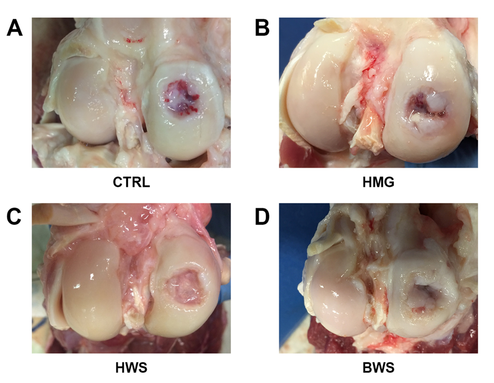
Development of a novel biphasic scaffold for osteochondral lesions

image description
Articular cartilage and subchondral bone frequently undergoes degeneration as a
result of traumas or disease. The osteochondral tissue has a poor healing potential, and current surgical procedures lead to the formation of a fibrocartilaginous tissue, which does exhibit reduced biomechanical properties. Due to the extremely different nature of cartilage and bone, modern approaches focus on the design and development of scaffolds combining distinct layers mimicking the morphological and mechanical heterogeneity.
Our Laboratory is involved in the development of a novel bi-phasic cellular/acellular scaffold for osteochondral regeneration. In collaboration with University of Salento, Politecnico di Milano, and University of Milan, we test in-vivo acellular scaffolds characterized by different materials and different architectures. The regenerative potential is analyzed by gross morphology studies, radiological images, biomechanical analysis and histochemical investigations of the osteochondral tissue itself and of the meniscus. Moreover, we investigate the chance of using scaffolds functionalized with chondrogenic or osteogenic molecules and/or seeded with different cellular populations.
Publications
- Peretti GM, Tessaro I, Montanari L, et al. Histological changes of the meniscus following an osteochondral lesion. J Biol Regul Homeost Agents. 2017; 31(4 Suppl 1): 129-134.
- Gervaso F, Mangiavini L, Di Giancamillo A, et al. Comparison of three novel biphasic scaffolds for one-stage treatment of osteochondral defects in a sheep model. J Biol Regul Homeost Agents. 2016; 30(4 Suppl 1): 24-31.
- Gobbi A, Scotti C, Karnatzikos G, Mudhigere A, Castro M, Peretti GM. One-step surgery with multipotent stem cells and Hyaluronan-based scaffold for the treatment of full-thickness chondral defects of the knee in patients older than 45 years. Knee Surg Sports Traumatol Arthrosc. 2017; 25(8): 2494-2501.
- De Girolamo L, Niada S, Arrigoni E, et al. Repair of osteochondral defects in the minipig model by OPF hydrogel loaded with adipose-derived mesenchymal stem cells. Regen Med. 2015; 10(2): 135-51.
- Sosio C, Di Giancamillo A, Deponti D, et al. Osteochondral repair by a novel interconnecting collagen-hydroxyapatite substitute: a large-animal study. Tissue Eng Part A. 2015; 21(3-4): 704-15.
- Deponti D, Di Giancamillo A, Gervaso F, et al. Collagen scaffold for cartilage tissue engineering: the benefit of fibrin glue and the proper culture time in an infant cartilage model. Tissue Eng Part A. 2014; 20(5-6): 1113-26.
- Marmotti A, Bruzzone M, Bonasia DE, et al. Autologous cartilage fragments in a composite scaffold for one stage osteochondral repair in a goat model. Eur Cell Mater. 2013; 26: 15-31.
- Gervaso F, Sannino A, Peretti GM. The biomaterialist’s task: scaffold biomaterials and fabrication technologies. Joints. 2014; 1(3): 130-7.
- Marmotti A, Bonasia DE, Bruzzone M, et al. Human cartilage fragments in a composite scaffold for single-stage cartilage repair: an in vitro study of the chondrocyte migration and the influence of TGF-β1 and G-CSF. Knee Surg Sports Traumatol Arthrosc. 2013; 21(8): 1819-33.
- Marmotti A, Bruzzone M, Bonasia DE, et al. One-step osteochondral repair with cartilage fragments in a composite scaffold. Knee Surg Sports Traumatol Arthrosc. 2012; 20(12): 2590-601.
